
Medicine occasionally encounters cases so extraordinary that they challenge everything doctors think they know about human development.
Most physicians will spend an entire career without seeing anything remotely similar. Yet every so often, a patient arrives whose condition expands the boundaries of medical knowledge.
One such case involved a newborn whose unusually enlarged head raised immediate concern.
Prenatal imaging had already revealed a large mass inside the baby’s skull. At first, specialists suspected a rare congenital abnormality, possibly a tumor. However, as additional scans were performed after birth, it became increasingly clear that this was something far more unusual.
The diagnosis would eventually place the infant among the rarest cases ever described in medical literature.
A Discovery That Defied Expectations
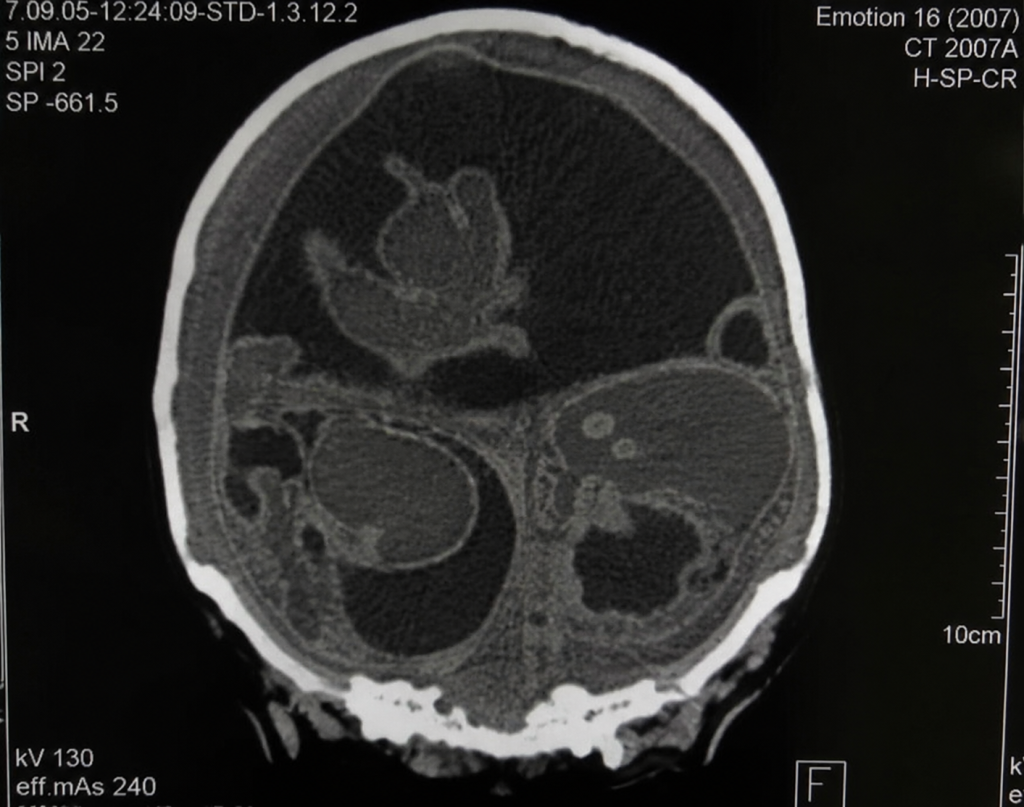
Detailed CT and MRI scans revealed not one abnormal mass but three separate structures occupying a significant portion of the cranial cavity.
These were not ordinary tumors.
Each contained partially developed fetal tissues arranged in a way that suggested early embryonic organization rather than random cell growth. Doctors identified developing bone, nervous tissue, skin, respiratory structures, and portions of the gastrointestinal tract.
For the medical team, the findings were astonishing.
Instead of a conventional congenital lesion, they were looking at an exceptionally rare developmental anomaly known as fetus-in-fetu.
What Is Fetus-in-Fetu?
Fetus-in-fetu is one of the rarest congenital conditions ever documented.
It occurs during the earliest stages of identical twin development, when one embryo becomes enclosed within the body of its sibling instead of continuing to develop independently.
Because the enclosed embryo depends entirely on the host’s blood supply, normal growth eventually stops. The result is a partially developed structure that may contain recognizable anatomical features but is incapable of surviving on its own.
Worldwide, only a few hundred cases have been reported.
Almost all of them involve a single parasitic twin located inside the abdomen.
Finding even one inside the skull is exceptionally uncommon.
Finding three in the same patient is almost unprecedented.
Why This Case Captured Worldwide Attention
The location alone made this case extraordinary.
The three fetal masses occupied valuable space inside the infant’s skull, placing increasing pressure on the developing brain.
Unlike the abdomen, where some fetus-in-fetu cases can remain undetected for months or even years, the rigid structure of the skull leaves virtually no room for expansion.
As pressure builds, the brain becomes compressed, creating an increasingly life-threatening situation.
Without intervention, long-term survival was considered highly unlikely.
Not a Tumor, but Something Even Rarer
The condition is often confused with teratomas, which are tumors capable of containing hair, teeth, bone, or other mature tissues.
Although both conditions may appear similar on medical imaging, they are fundamentally different.
Teratomas consist of disorganized collections of multiple tissue types.
Fetus-in-fetu, however, demonstrates recognizable embryonic organization. In some reported cases, doctors have identified primitive spinal structures, limb buds, or other anatomical arrangements that resemble early fetal development.
These distinctive features are what ultimately led specialists to classify the intracranial masses as fetus-in-fetu rather than a congenital tumor.
An Extremely High-Risk Operation
As the infant’s condition worsened, surgery became the only possible treatment.
The procedure presented enormous challenges.
Operating inside the skull of a two-month-old child is already one of the most demanding tasks in pediatric neurosurgery. Removing three complex masses while protecting the surrounding brain tissue increased the difficulty even further.
Every surgical decision carried significant risk.
Every movement had the potential to affect the child’s survival.
Despite the efforts of the multidisciplinary medical team, tragedy followed.
During the operation, the infant suffered cardiac arrest.
Resuscitation attempts were unsuccessful, and the child could not be saved.
What Researchers Learned
Although heartbreaking, the case provided valuable insight into one of medicine’s least understood developmental abnormalities.
Genetic analysis demonstrated that the infant and the three enclosed fetal structures shared the same genetic profile, strongly supporting the theory that fetus-in-fetu originates from abnormal development during an identical twin pregnancy rather than representing a true tumor.
Even today, researchers continue to investigate the precise biological mechanisms responsible for this phenomenon.
Because the condition is so exceptionally rare, every documented case contributes important information that may improve diagnosis and understanding in the future.
A Reminder of How Complex Human Development Truly Is
Human embryonic development is an astonishingly precise process.
Within just a few weeks after conception, billions of cells must divide, migrate, and organize with remarkable accuracy to form every organ and structure in the body.
In extraordinarily rare circumstances, a small disruption during these early stages can lead to developmental anomalies unlike anything most physicians will ever encounter.
The case of this infant remains one of those remarkable exceptions.
It stands as both a profound medical mystery and a reminder that, despite decades of scientific progress, human development still holds secrets that modern medicine has yet to fully explain.
Behind the scientific significance lies the story of a family who hoped surgery would offer their child a chance at life. While that hope ultimately ended in tragedy, the case continues to help researchers better understand one of the rarest congenital conditions ever recorded.