
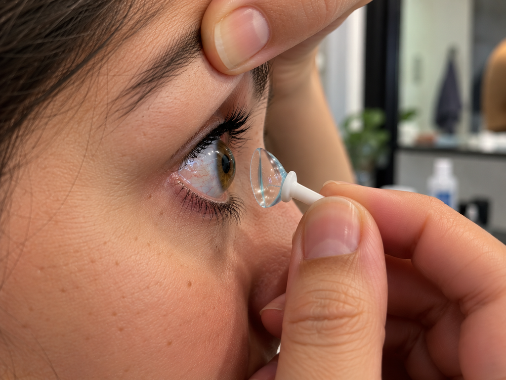
Watching someone remove a scleral contact lens for the first time can be unsettling. The lens looks unusually large, the eye appears to move slightly as it is removed, and a tiny suction tool may make the whole procedure look far more aggressive than anything associated with ordinary contact lenses.
Yet the dramatic appearance hides a sophisticated piece of medical technology.
Scleral lenses are not simply oversized versions of conventional contacts. Their shape, the way they interact with the eye, and the conditions they can help manage are fundamentally different. For some people with complex corneal disease, they can provide a level of vision and comfort that glasses or standard contact lenses cannot.
A contact lens designed to vault over the cornea
Most people think of a contact lens as something that sits directly on the transparent cornea at the front of the eye. A scleral lens works differently.
It is a large-diameter, rigid gas-permeable lens designed to vault over the cornea and rest on the sclera, the white part of the eye. Before insertion, the bowl of the lens is filled with preservative-free sterile saline solution.
Once the lens is in position, a reservoir of fluid remains between its inner surface and the cornea.
That liquid layer is one of the defining features of scleral lenses. Instead of forcing an irregular cornea to conform to a lens, the system essentially creates a new, smooth optical surface in front of it. Medical literature describes this fluid reservoir as an important part of both the optical and therapeutic function of scleral lenses.
Why would someone need such a large lens?
One of the best-known reasons is keratoconus.
In a healthy eye, the cornea has a relatively regular curvature. In keratoconus, the cornea progressively thins and protrudes outward, developing a more cone-like and irregular shape. That distortion can produce blurred or distorted vision that becomes increasingly difficult to correct with ordinary glasses.
Because a scleral lens vaults over the irregular cornea, the fluid trapped underneath helps neutralize some of those irregularities. The front surface of the lens then provides a more regular refractive surface through which light can enter the eye.
This is not merely theoretical. Research involving people with keratoconus has found substantial improvements in visual acuity and vision-related quality of life with scleral lenses, although researchers also emphasize that larger and better-controlled studies are still needed.
But keratoconus is only part of the story.
Scleral lenses may also be prescribed for other forms of corneal irregularity, including some post-surgical corneas and corneal scarring. They can additionally play a therapeutic role in certain severe ocular-surface diseases.
The tiny reservoir that changes everything
There is another reason these lenses are unusual.
The saline reservoir beneath the lens continuously bathes the corneal surface while the lens is being worn. For patients with certain severe forms of ocular surface disease, that protected environment can be particularly valuable.
This is one reason scleral lenses have been investigated and used for severe dry-eye conditions when conventional approaches are insufficient. However, not every person with dry eyes needs — or would benefit from — scleral lenses, and the evidence varies depending on the underlying condition.
They are specialized medical devices, not a universal upgrade from ordinary contacts.
Scleral lenses, soft lenses and conventional rigid lenses are not the same thing
A soft contact lens generally drapes over the cornea and moves slightly with blinking. A traditional corneal rigid gas-permeable lens is much smaller and sits primarily on the cornea.
A scleral lens takes another approach entirely: it spans the cornea and lands farther out on the ocular surface.
That difference explains its distinctive appearance. It also explains why fitting one requires considerably more than simply choosing the correct prescription.
The specialist must consider the shape of the eye, how much clearance exists between the lens and cornea, how the lens lands on the scleral region and how the tissues behave after hours of wear. Oxygen reaching the cornea is another important consideration.
In other words, two people with identical spectacle prescriptions could require very different scleral-lens designs.
Why removal can look surprisingly aggressive
This is the part that often attracts attention on social media.
After a scleral lens has been worn for hours, it can form a relatively stable seal against the ocular surface. Simply grabbing the lens and pulling outward is therefore not the goal.
Many wearers use a small suction device, often called a removal plunger. The important detail is where it is placed.
Guidance from the Scleral Lens Education Society instructs users to attach the removal device toward the lower edge of the lens rather than directly in its center. The lens can then be tilted outward, helping break the seal before it is removed.
That distinction explains why a removal technique shown online can look uncomfortable or even alarming. The objective should not be to overpower the suction by pulling harder. It is to release the edge of the lens so the seal is broken.
Scleral Lens Education Society handling guide
Why proper technique matters
The eye is not simply a loose sphere sitting inside the socket, so routine scleral-lens removal is not pulling the eyeball out of position in the way a dramatic video might suggest.
But that does not mean technique is irrelevant.
Incorrect handling can cause discomfort or irritation, and scleral-lens users can encounter other problems such as difficulty inserting or removing the lenses, debris accumulating in the fluid reservoir, fogging, corneal swelling and problems related to an inappropriate fit.
As with other contact lenses, hygiene is also essential. Contact-lens-related microbial keratitis is uncommon but potentially serious, which is why cleaning, disinfection, handling and the solutions used with the lenses matter.
Persistent pain, significant redness, light sensitivity, discharge or a sudden deterioration in vision while wearing contact lenses warrants prompt professional assessment rather than simply continuing to wear the lens.
The lens is large for a reason
There is an interesting contradiction at the heart of scleral lenses.
Their size is precisely what makes them look intimidating, yet their size is also what allows them to perform their unusual function.
Rather than sitting on a damaged or highly irregular cornea, the lens can bridge over it. The saline underneath fills the space between the cornea and the lens, while the rigid front surface creates a much more regular optical interface.
A 2025 systematic review examining scleral-lens use in keratoconus included 463 eyes and reported consistent improvements in visual acuity and vision-related quality of life. The authors also noted limitations in the available research and called for stronger controlled studies, an important reminder that promising clinical results should not be turned into exaggerated claims.
That context changes the way these viral videos look.
What initially resembles an enormous contact lens being suctioned off someone’s eye is actually the final step in using a carefully fitted optical device. For many wearers, inserting and removing it eventually becomes an ordinary daily routine.
And behind that strange-looking piece of transparent material is a remarkably elegant idea: when the cornea itself can no longer provide a smooth optical surface, it may be possible to create a new one just above it.